Answer: The substantia nigra is a midbrain structure that is part of the basal ganglia and is important for movement.
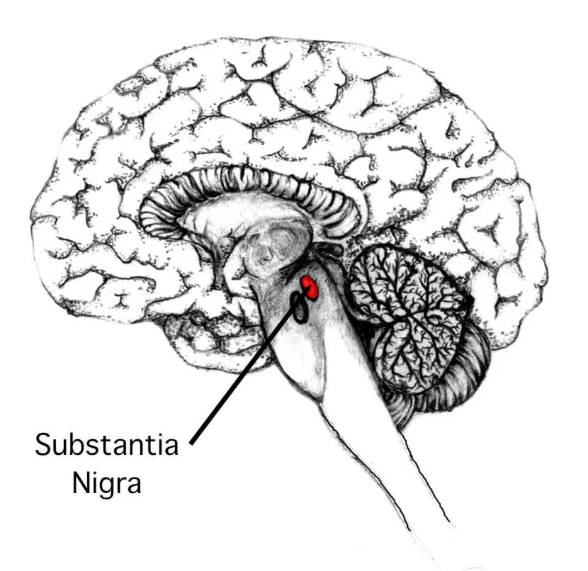
The substantia nigra is a brain structure located in the midbrain. It is part of a series of interconnected structures called the basal ganglia. The substantia nigra is the largest nucleus of the midbrain, and we have one in each hemisphere.
The healthy substantia nigra is visible in an MRI scan in an axial slice. It is sometimes called a “swallow tail sign” due to the twin pronged appearance, with one in each hemisphere.
Although the substantia nigra is sometimes considered to be a single brain structure, it is more accurately divided into two different regions, called the substantia nigra pars compacta (SNc) and substantia nigra pars reticulata (SNr). The regions are divided this way since each of the two receives and sends different connections and carry out different roles within the circuitry of the brain.
Substantia nigra pars reticulata (SNr)
The reticulata receives input connections from the striatum, two GABA-ergic inputs from the direct and the indirect pathway. The direct pathway neurons are striatal medium spiny neurons (spiny projection neurons) that express the dopamine D1 receptors. These direct pathway neurons inhibit the activity and output of the SNr.
This area also receives striatal input through the indirect pathway. The GABA-ergic output projects to the external capsule of the globus pallidus. Those cells then project to the subthalamic nucleus, which contain glutamatergic projections into the SNr.
The reticulata contains many GABA-ergic inhibitory neurons that are spontaneously active. On average, these neurons fire very rapidly at a rate of 25 Hz. The major outputs of the reticulata project to the thalamus and the superior colliculus.
The cytology of this structure is very similar to the internal capsule of the globus pallidus.
Substantia nigra pars compacta (SNc)
Neurons in the compacta are responsible for motor control. Within this area are a large population of dopamine producing and releasing neurons. These dopaminergic cells in the substantia nigra are often darker than the surrounding cells, which is how the region got named. These cells have a high concentration of a pigment called neuromelanin, and this causes them to appear dark.
These dopaminergic neurons are the ones that are particularly sensitive to destruction that lead to the onset of Parkinson’s disease. Usually, by the time a person presents to the clinic with symptoms of PD, they have already lost the majority of cells in the substantia nigra pars compacta.