Answer: An arteriovenous malformation (AVM) is a structural anomaly of the circulatory system that may lead to a stroke.
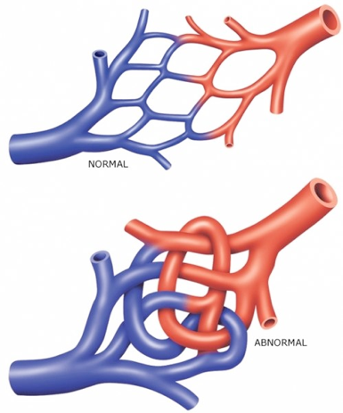
In a typical average brain, a dense network of tiny blood vessels called the capillaries exists at the junction between the arteries and veins. The capillaries are highly efficient structures designed to pass oxygenated blood into the neighboring cells. The capillaries are also designed to buffer the difference in blood pressure, which is generally very high in arteries but very low in the veins. The blood vessel walls of capillaries are very thin, which allows the exchange of air: oxygen permeates out of the capillaries, and carbon dioxide permeates into the capillaries.
However, in an arteriovenous malformation, a person may have fewer capillaries in one of the junctions. Instead, they may have a direct connection between the artery and vein. These are generally observed in the central nervous system, especially in the brain, but may still develop outside of the nervous system. In most cases, an arteriovenous malformation is harmless, but it can lead to complications including headaches, seizures, or more severely, strokes. They are very rare, affecting only less than 1% of people. In the US, the prevalence is estimated to be one in 200,000 people. It is believed that they form while young during development. Symptoms may develop when the patient is in their teenage years up until their 40s, but can manifest beyond this. However, in general, once a person is in their middle age, the AVM becomes more stable, and generally does not present with any symptoms.
The symptoms of an arteriovenous malformation are not the same across individuals. Instead, the symptoms differ based on where the malformation is located.
In an arteriovenous malformation, the capillary system is missing and unable to buffer the high pressure incoming from the heart. As a result, the junction between the artery and the vein experiences tremendous pressure, which can cause the blood vessels to expand outwards. This expansion of the AVM can strain or damage the walls of the blood vessels, making them prone to injury. Arteriovenous malformations may lead to a stroke, which is a bursting of blood vessels in the brain, leading to uncontrolled blood spillage in the brain. The uncontrolled blood flow causes severe damage to brain cells. There are three types of strokes, with the majority of them being either ischemic or hemorrhagic stroke. A small fraction of strokes (2-3% of strokes) are because of a rupture in an arteriovenous malformation. Most people with AVMs do not experience any symptoms until a hemorrhage occurs as a result of the AVM rupturing.
Many arteriovenous malformations (AVMs) are congenital - people are often born with them. But in some bizarre cases, they can develop as a person gets older. It is also unclear as to why people are born with them, in some cases it is genetic but not always. It is estimated that there is a 2% annual yearly risk of an AVM that leaks, causing a hemorrhage which leaks blood into the brain.
Another consequence of an AVM is that the brain may be deprived of oxygen compared to a person with a normal capillary bed between their arteries and veins in the brain. Because the capillary bed is designed for sending oxygenated blood into every part of the brain, with an AVM the brain may experience relatively low levels of oxygen. In the AVM, the blood is flowing too quickly for the adjacent neurons to take up oxygen and to pass off carbon dioxide. Whereas this is not explicitly the dangerous hypoxia that might result from a stroke, small decreases in oxygen flow to the brain as a result of poor diffusion may have subclinical consequences.

In a severe form of an AVM called the “vein of Galen” malformation, an arteriovenous malformation is discovered early in life. The symptoms generally appear soon after birth. Often times as a result of the vein of Galen defect, the blood fluid balance is disrupted, resulting in an increase in the fluid inside the brain. This may result in a form of hydrocephalus, causing the head to swell, as the younger skull is not fully fused. These swollen veins may result in early symptoms such as swollen veins that are visible, congestive heart complications, and seizures. All of these symptoms resulting from the vein of Galen defect should be treated immediately.
There are five different patterns, or types of vein of Galen malformations. A pattern 1 malformation is when multiple blood vessels, such as the anterior and superior cerebral arteries and thalamic perforating arteries all drain into the vein of Galen. A pattern 2 malformation is when one posterior choroidal artery connects directly into the vein of Galen. A pattern 3 malformation is when the posterior choroidal artery and the anterior cerebral arteries make a direct connection into the vein of Galen. The pattern 4 malformation is defined by a series of complex vessels consisting of the posterior choroidal and thalamic perforating arteries send blood directly into the vein of Galen. In a pattern 5 malformation, arteries in the right inferior frontal lobe sends blood directly through the inferior sagittal sinus into the vein of Galen.
Many of these vein of Galen malformations leads to severe complications, with about 70% of cases leading to mortality. Even after surgical repair of the arteriovenous malformation, the mortality rate remains high, almost 40%.
Arteriovenous malformations can develop in anyone. The rate of incidence tends to be higher in males compared to females. Also, having a family history of AVMs may be a contributing factor, although the genetics of the disorder have not been fully worked out.
A ruptured arteriovenous malformation resulting in a stroke was described by the neuroscientist Dr. Jill Bolte Taylor in her nonfiction first hand account of a stroke, My Stroke of Insight.