Answer: Endolymph is the fluid that is inside the membranous labyrinth of the inner ear.
The inner ear is a complex organ that is responsible for such functions as auditory sensation (hearing) and the vestibular system (balance and spatial orientation.) Both of these senses rely on specialized sense organs such as the cochlea for auditory sensation and the semicircular canals and otoliths vestibular sensation. For these sensory systems to work properly, a fluid called endolymph is used.
Endolymph is essentially the extracellular solution for the nerve cells in the auditory and vestibular systems. Endolymph itself is mostly water, but it has a high concentration of solutes dissolved in it. The main cation in endolymph is potassium, which is at 150 mM. Sodium is also present at 1 mM. This difference makes endolymph different from the normal extracellular solution that is found surrounding most nerve cells. In neurons bathed in endolymph, positively-charged potassium ions are the main charge carrier during depolarization. On the other hand, for neurons surrounded by the normal cerebrospinal fluid, positively-charged sodium ions are the charge carrier during depolarization.
Auditory system
The endolymph in the auditory system is the fluid that helps convey a physical stimulus, the compression and rarefaction of air waves, into an electrical and chemical signal that the brain can interpret. The endolymph is the interface that transmits the information from the air waves into the inner ear.
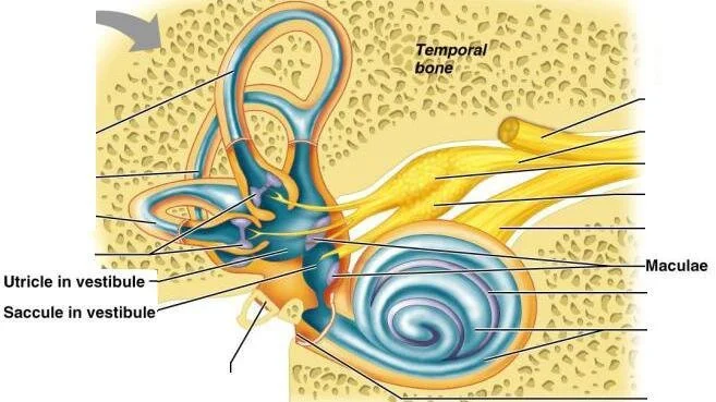
The oval window is a thin membrane that serves as the boundary between the middle ear, which is surrounded by air, and the cochlea of the inner ear, which is surrounded by endolymph. Airwaves are detected by the eardrum, which is the membrane that separates the outer ear from the middle ear. Within the middle ear are three tiny bones called ossicles that then transmit air wave information through the oval window into the endolymph of the inner ear.
Within the inner ear, lining the inner surface of the cochlea are several neuronal cells called hair cells. These are named because their appearance. Hair cells have long protrusions called stereocilia, which stick into the endolymph that fills the inside of the cochlea. The endolymph here vibrates at the same frequency as the air when sound waves are detected, and this vibration information is sensed by the stereocilia. These hair cells contain special ion channel proteins on the cell surface that are sensitive to mechanical deflection. When a sound wave passes over a hair cell, these ion channels open, which allows potassium to enter the neurons. Unlike the rest of the nervous system, this potassium causes depolarization, and that excites the cell.
These hair cells communicate with the brain by sending action potentials down the auditory nerve, one branch of the vestibulocochlear nerve (Cranial Nerve VIII).
Vestibular system
The vestibular is our sense that helps us figure out which direction is up and down, and also helps us process changes in head acceleration. These sensory functions require two organs in the inner ear, the semicircular canals and the otoliths, respectively.
The semicircular canals are a series of three membranous tubes, one in each of the three dimensions. The canals are filled with endolymph, and just like the cochlea, have hair cells with stereocilia that extend into the endolymph. These hair cells are found at the end of each of the canals, in an expansion called the ampulla. When the head moves in one direction, the fluid moves as well, causing the hair cells to deflect. When the hair cells move in one direction, mechanically gated ion channels will open, allowing potassium ions to enter the cell, resulting in depolarization. But, when they move in the opposite direction, these channels close, resulting in hyperpolarization. Collectively, these signals convey head movement and tilt information to the brain via the vestibulocochlear nerve (Cranial Nerve VIII).