Answer: The loss of dopamine in Parkinson's disease results in decreased direct pathway output activity.
The major pathological sign of Parkinson's disease is the loss of dopamine producing neurons in the substantia nigra pars compacta that send projections into the striatum. When these neurons die, there is a decrease in the amount of dopamine in the striatum. Because dopamine functions to allow regular motor output, decreasing dopamine produces the symptoms of Parkinson’s disease, including bradykinesia and difficulty with movement.
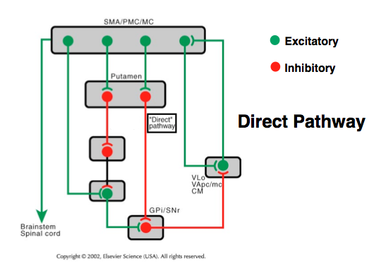
In the striatum, there are several types of cells. The most prevalent are the medium spiny neurons, or spiny projection neurons. They make up about 95% of striatum neurons in the rodent brain. These cells possess one of two types of dopamine receptors, either the excitatory D1 or the inhibitory D2 receptors. The D1 expressing medium sized spiny neurons make up the direct pathway, while the D2 expressing MSNs make up the indirect pathway.
The D1 expressing medium spiny neurons send their inhibitory GABA-ergic and substance P producing axons into a region of the brain called the substantia nigra pars reticulata (SNpr) and the globus pallidus internus. This is called the striatonigral projection. These cells then project to and inhibit the thalamus. Finally, the thalamus excites the motor cortex.
In a healthy brain, dopamine release activates the D1 receptors, which excited the output of the direct pathway. This activation encourages motor output, through the normal activity of the thalamus. However, in the Parkinson's brain, the loss of dopamine means a decrease in activity.