Answer: The circumventricular organs are a group of brain structures that surround the third and fourth ventricles and play a role in either sensing signals from the body, or sending signals to the body.

The brain collects information from the outside world using sense organs, such as the eyes and ears. However, the brain also has to monitor the internal state of the body. To do this, the brain relies on a group of organs that are clustered around the third and fourth ventricles in the brain. Additionally, the brain needs to send signals to the rest of the body via release of neurohormones into the bloodstream. The brain structures that carry out these functions are sometimes collectively called the “circumventricular organs”.
The major reason these organs are able to carry out these sensory or secretory functions is due to their anatomy. All organs in the body must receive oxygen to function. That oxygen is carried to the organs by the circulatory system as the blood is transported throughout the body. In the brain, there is a structure that surrounds each blood vessel called the “blood-brain barrier.” The purpose of the blood brain barrier is to separate toxic substances from the blood from getting into the brain through the formation of tight junctions across glia. In the blood vessels that innervate the brain structures near the circumventricular organs, the blood brain barrier is more permeable than in other brain structures. The increased permeability means that these organs are able to sense significant changes in blood chemistry and respond appropriately.
Circumventricular organs, or CVOs, can be classified into two major groups based on their primary function: sensory or secretory.
Circumventricular organs with a sensory function:
Area Postrema
The area postrema is specific for identifying if there is an unusually high concentration of toxins in the blood and cerebrospinal fluid that derived from substances in the stomach. Area postrema is located in the medulla and is responsible for triggering the vomiting reflex in the event that the toxins in the blood that may cause some kind of physical damage to the body.
Area postrema is located at the very base of the fourth ventricle. The cells in the area postrema are not neuronal in nature, but rather glial cells. Area postrema is made up of ependymal cells which lack tight junctions and therefore makes a direct interface with the blood, making area postrema responsive to physiological changes.
Subfornical Organ
The subfornical organ functions in regulating the amount of water in the blood. It can do this by communicating with the kidneys through the hormonal release of vasopressin into the bloodstream. Cells in the subfornical region also have receptors for several different neurohormones.
Anatomically, the subfornical organ is divided into two main groups of cells, the central zone and the caudal / rostral zones. The central zone is highly vascularized with capillaries, and consists of glial cells and neuronal cells. On the other hand, the caudal and rostral regions are less densely vascularized and consists of mostly of nerve fibers rather than cell bodies.
Vascular Organ of Lamina Terminalis
The vascular organ of the lamina terminalis serves a function in maintaining the proper osmolarity balance in the blood. It receives inputs from the brain stem and hypothalamus. It is located at the base of the third ventricle.
Circumventricular organs with a secretory function:
Pituitary gland
The pituitary gland serves a wide variety of endocrine functions by releasing hormones directly into the bloodstream. It is often considered the “master gland” of the body since the hormones released carry out such a wide variety of physiological functions towards making the body function. The pituitary gland can be separated into two major anatomical divisions, the anterior (adenohypophysis) and the posterior (neurohypophysis) pituitary gland, each of which carries different functions. The anterior pituitary is non-neural; all the cells in this area are secretory. The anterior pituitary releases such hormones as adrenocorticotropic hormone, thyroid-stimulating hormone, and growth hormone for example. The posterior pituitary is made up primarily of axonal projections that originate in the hypothalamus. The anatomical tract that sends signals from the hypothalamus is called the infundibulum. The two major neurohormones that are stored in the posterior pituitary are oxytocin and vasopressin.
Pineal gland
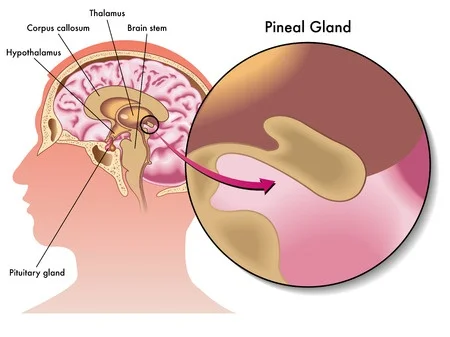
The pineal gland is important for the release of melatonin into the bloodstream. Melatonin is a signaling hormone that triggers sleepiness and naturally rises at the end of the day. The suprachiasmatic nucleus receives inputs from the eyes via the retinohypothalamic tract, and regularly suppresses suprachiasmatic nucleus activity in the day time. At night there is low activation of the suprachiasmatic nucleus, which relieves the inhibition on the suprachiasmatic nucleus, resulting in elevated melatonin release into the blood.
The pineal gland also may store a variety of peptides that get secreted into the blood, including vasopressin, oxytocin, neuropeptide Y, and substance P. Vasopressin is a kidney related signal that helps maintain the correct osmolarity in the blood, and may be involved in feelings of romantic love. Oxytocin and vasopressin are both chemical signals that are both important for attraction and feelings of romantic love. Neuropeptide Y, also called NPY, is the most common peptide that can be found in the central nervous system. NPY functions in guiding neurogenesis. Substance P is an 11-chain peptide that is involved with inflammation and pain processing by the brain.
The pineal gland and pituitary gland are often confused for one another.
Subcommissural organ
The subcommissural organ lies at the entrance to the cerebral aqueduct and is made up entirely of ependymal cells rather than neurons. It’s function is not very well characterized by researchers, but a few features have been identified. It produces a glycoprotein signaling molecule called SCO-spondin, which is related to maintaining the structural features of the cerebral aqueduct. If there was an injury to the subcommissural organ, it could result in a failure for the aqueduct to remain open, resulting in a potentially deadly condition called hydrocephalus.
Median eminence
The median eminence is made up of tanycytes, which are highly specialized types of ependymal cells. It is involved with the release of gonadotropin releasing hormone into the bloodstream. Anatomically, it is located within the hypothalamus.