Answer: The uncinate fasciculus is a white matter tract in the brain that connects the anterior temporal lobe and the frontal lobes of the cortex.
In the nervous system, the color of the tissue gives information about the function of those areas of the brain. Gray matter contains mostly the cell bodies of neurons. On the other hand, white matter contains the axons of those neurons. White matter structures are mostly used for communicating information between major circuits of neurons.
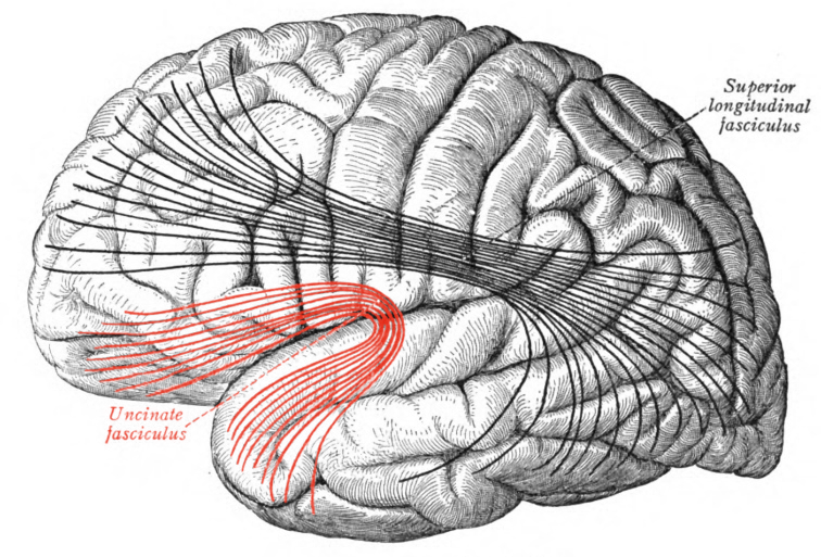
Structure of the uncinate fasciculus
The uncinate fasciculus ia a long white matter tract. The fiber tract has one end in the pole of the temporal lobe, which is the part of the cortex behind the ears. Here, it is connected with many of the limbic structures, parts of the brain that are strongly involved with our processing of emotional states and memories. Some limbic structures connected with the uncinate fasciculus include the amygdala and parahippocampal gyrus.
From there, the uncinate fasciculus curves upwards and anteriorly towards the frontal lobe. In the frontal lobe, the white matter fibers connect with other important structures like orbitofrontal cortex, one part of the prefrontal cortex. The orbitofrontal cortex is important when it comes to cognitive flexibility and learning appropriate responses.
The uncinate fasciculus is a bi-directional pathways, which means that axons are sent outwards from the temporal lobe and out of the frontal lobe.
The precise shape and angle of the uncinate fasciculus can be determined using an imaging method based on fMRI technology called diffusion tractography, or DTI. In DTI, the movement of water molecules are tracked, and this is used to differentiate white matter from gray matter.
Function of the uncinate fasciculus
The uncinate fasciculus is important for a wide range of congitive and emotional functions. Some functions include:
Memory. The parahippocampal areas are important structures for the creation of memory related circuits, which are communicated towards the orbitofrontal cortex for more longer term storage. One specific type of memory attributed to uncinate fasciculus is reversal learning. In an example reversal learning experiment, a person may learn to push a button whenever they see a green stimulus to get a reward. After several successful trials, the person may need to NOT push the button when they see the green stimulus to get the reward.
Emotional regulation. The circuits of the amygdala are important for the formation of emotionally related memories, such as creating associations between innocuous stimuli and pain. The related frontal lobe circuits are involved in impulse control and inhibition of behaviors. Being able to control an emotional outburst during distressing situations, for example, is a behavior that the uncinate fasciculus likely mediates.
Language. The inferior frontal gyrus (IFG) is a brain structure that has a strong role in the production of language. Within the IFG is an area called Broca’s area, which when damaged, produces a language disorder called expressive aphasia. Connections between the IFG and temporal lobe through the uncinate fasciculus is probably important for language related memories, such as when a person is learning new words.
Clinical implications of the uncinate fasciculus
Autism. Among verbal autistic people (diagnosis of autism spectrum disorder), there is a difference in uncinate fasciculus volume between the left and right hemispheres, while no hemispheric differences are found in either typically developing people or nonverbal autistic people. (Structural connectivity in ventral language pathways characterizes non-verbal autism)
Major depressive disorder. Brain imaging strategies have shown that decreased left uncinate fasciculus volume is correlated with major depressive disorder. (Reduced myelin density in unmedicated major depressive disorder: An inhomogeneous magnetization transfer MRI study)
Alzheimer’s disease. Using diffusion tensor imaging, it was discovered that the uncinate fasciculus had lower fractional anisotropy in patients with Alzheimer’s disease compared to age matched control participants. Fractional anisotropy is a measure of white matter degeneration. (Diffusion abnormalities of the uncinate fasciculus in Alzheimer’s disease: diffusion tensor tract-specific analysis using a new method to measure the core of the tract)