Answer: The Brodmann areas were an early method by which brain regions were divided based on the types of cells contained within.
By Mark Dow. Research Assistant Brain Development Lab, University of Oregon. [Public domain], via Wikimedia Commons
Korbinian Brodmann was a German neuroanatomist in the late 1800s and early 1900s. He is best known for dividing the cerebral cortex of the brain into 52 different regions based on their histological properties.
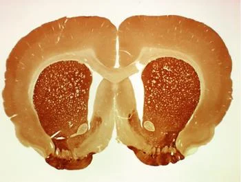
His major study in cytoarchitecture was published in 1909 (Comparative Localization Studies in the Brain Cortex, its Fundamentals Represented on the Basis of its Cellular Architecture). In this work, he analyzed the brains of humans, nonhuman primates, and other species (keep in mind that having the same Brodmann numbers across species does not indicate that the region of the brain performs the same function.) By taking different parts of brains and processing them with a Nissl stain, he could examine the types of cells contained within. He looked at general cytoarchitecture as well as internal circuitry of how cells were connected.
One of Brodmann’s theories was that each of these different parts of the brain performed a different function, a line of thought that still persists today.
As of present times, the general consensus in the field of neuroscience is that the Brodmann numbers are still a helpful approximation of different brain regions. The activation of parts of the brain as studied with functional imaging such as fMRI or PET scanning often closely correspond to activation of specific Brodmann areas. There is some overlap between the functions of brain areas, so the Brodmann areas serve only as an approximation rather than a boundary. However, most scientists refer to regions of the brain by a specific name rather than the Brodmann number.
Here are some particularly interesting Brodmann regions and their function.
- Brodmann area 1, 2, 3: Primary somatosensory cortex, which processes tactile sensory and proprioceptive information
- Brodmann area 17: Primary visual cortex (V1), which processes visual information as received by the retina.
- Brodmann area 37: Fusiform face area, responsible for processing of facial information. Injury to this area may result in prosopagnosia, an inability to see faces.
- Brodmann 41, 42: Primary auditory cortex (A1), which receives sound information through the ears.
- Brodmann 43: Gustatory cortex, which processes taste information from the mouth.
- Brodmann area 44, 45: Broca’s area, in the left hemisphere, is responsible for speech comprehension and production.
![By Mark Dow. Research Assistant Brain Development Lab, University of Oregon. [Public domain], via Wikimedia Commons](https://images.squarespace-cdn.com/content/v1/5a96f42d5b409bfd5be103ca/1525793354846-RZRZTMBEHQ4YARFWEZHQ/brodmann+areas+history)


