In studying neuroanatomy, it is useful for the field if a unified set of language is used to refer to different structures of the nervous system. This way, ambiguous terms like “top” or “front” can be eliminated.
(Note: all of these terms refer to a person if they were lying down on the floor, with their chin on the ground, looking forward in the direction of their body.)
Brain and spinal cord (central nervous system)
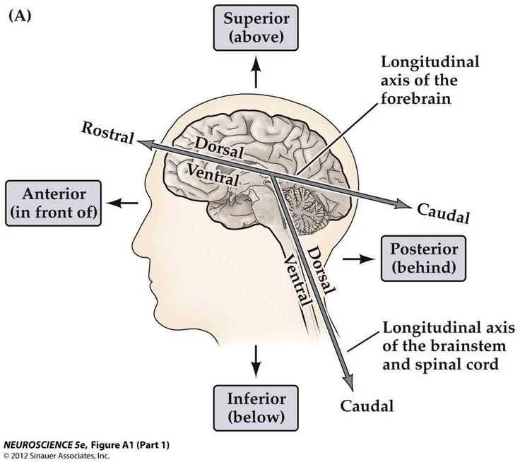
The first pair of terms in neuroanatomy is to distinguish the top of the brain from the bottom of the brain. In neuroanatomy, we use the word dorsal to refer to structures above, closest to the skull surface. On the other hand, we use ventral to describe structures at the base of the brain, closer to the chin or neck. “Dorsal” originates from the late Latin or middle French word dorsualis, which means “of or pertaining to the back.” When a person is lying down on the ground, the top of their skull is the same side as their back. “Ventral” originates from French or Latin, and means “of or pertaining to the belly.”
Sometimes, structures are referred to by the words “superior” or “inferior”. These words correspond with dorsal and ventral, respectively. For example, a structure that is superior to another structure is located dorsal to it.
The next axis is the “front-to-back” direction, going along the direction from the nose to the back of the head. In this plane, the front is rostral and the back is caudal. Rostral comes from rostrum, meaning “nose”. Caudal comes from the word which means “tail”. Another pair of words that correspond to this direction is anterior and posterior. The best way to remember this is to think of a person lying down, looking forward parallel across the floor. Brain structures that are caudal (posterior) are closer to the “tail” of the body: the cocyx, or the end of the spinal cord near the butt.
The last set of neuroanatomical terms refer to the “side-to-side” direction, from ear to ear. Instead of a left versus right direction, neuroanatomists think of this plane as middle versus side. To make the distinction, they would say that a structure in the middle is medial and a structure to the side is lateral. In using lateral, they do not distinguish between left and right hemispheres, largely because many (but not all) brain structures are symmetrical across the midline.
In the spinal cord, structures use the same set of language. For example, a spinal cord structure that is closer to the back is dorsal, while one that is closer to the front of the body is ventral. Structures that are closer to the center of the spinal cord are medial compared to structures that are closer to the sides, which is described by the word lateral. Since almost all images of the spinal cord are really taken in this orientation, a since section of spinal cord will not always contain significant tissue in the anterior or posterior direction.
Peripheral nervous system
Outside of the brain and spinal cord, there are several nerves that innervate other organs, such as internal organs as well as the muscles that control our skeletal system. To describe these nerves, neuroanatomists use words such as proximal and distal. A nerve that innervates tissue or receives information from tissue that is closer to the central nervous system is proximal. On the other hand, nerves that innervate tissue or receive sensory information from tissue farther from the central nervous system is distal. For example, the nerves projecting to and from the skin at the shoulder are proximal than the nerves that project to and receive information from the fingertips.