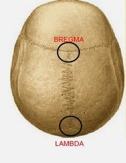
Answer: In neuroanatomy, bregma and lambda are two locations on the surface of the skull that allows for stereotactic identification of parts of the brain.
In neurosurgery or in research, it is important to know where in the brain a surgical intervention will take place. Ideally, the skull should remain as together as possible, so drilling a small hole is preferable. We use a set of three dimensional coordinates in order to know where the hole should be drilled.
These coordinates rely on anatomical markers that are uniform across individuals. There are two major anatomical markers on the dorsal surface of the brain that are formed when the plates of the skull fuse during development, and these markers are used to identify the location of various anatomical structures of the brain.
The anterior most marker is called bregma. Bregma is the spot where three cranial plates, the frontal bone (behind the forehead) and the two parietal bones (the top sides of the head) meet. You can identify bregma because it is the intersection of the two sutures, the coronal suture and the sagittal suture. The word bregma is of Greek origin, meaning "top of the head."
The more posterior marker is called lambda. Lambda is identified as the spot where the three cranial plates, the two parietal bones and the occipital bone (back of the head) meets. Lambda is the upside-down, broad v-shaped point that is indicated by the intersection between the sagittal suture and curved lambdoid suture.
(However, some research groups use the extrapolation of the lambdoid suture with the sagittal suture as lambda. This point is more posterior than the intersection of the three plates. These research groups call the point of three intersections “true lambda.” There is not a strong consensus in the field which is the correct location of lambda.)
Bregma and lambda in research
These anatomical markers are important in research. It is common for the scientist to perform brain surgery with the help of a tool called a stereotax, which is essentially a frame that fits around the skull. Stereotaxic frames have been developed for non humans as well, including frames for rats or mice. In conjunction with a brain atlas, like one hosted at the Allen Institute, the scientist can precisely pinpoint the location of different brain structures relative to bregma or lambda.
In an adult (postnatal day 60+) C57/B6 mouse, the average distance between bregma and lambda is around 4.2 millimeters.


