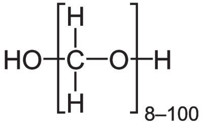
Answer: Paraformaldehyde (PFA) is a chemical fixation agent that preserves tissue by forming covalent bond cross links between molecules.
In order to analyze biological tissue on a molecular scale, a fixative compound is often used. The tissue is exposed to the fixative agent, which functions by cross linking the components of the tissue, essentially “locking” them in place and time.
In order to analyze the intracellular components, the cell membranes must be permeabilized. This means that tiny microscopic holes must be punched in the cell membrane, which allows antibodies and dyed to enter into the cell membrane. If the tissue were not fixed at this stage in the process, the permeabilization agent would completely destroy the tissue, and cellular material would be washed away entirely. The shapes of the cell membranes wouldn't be preserved.
Another advantage of fixing tissue is to lock the material in time, preventing further degradation of the tissue. Once biological tissue is deprived of blood flow, it loses nutrients and oxygen, both of which are essential for normal function. This is part of the process of cell death. When biological tissue is fixed, the covalent cross links prevent cell death cellular cascades from functioning, resulting in a “frozen” sample.
Paraformaldehyde may be introduced to the sample in one of two main methods.
First, the entire body can be perfused with fixative. In this process, a thoracotomy is performed on a living, anesthetized non human animal. This procedure gives the experimenter access to the heart. The PFA solution is pumped via a syringe directly into the left ventricle, which is the major pump of the heart. The heart proceeds to pump the fix throughout the circulatory system. An incision is also made in the right atrium to relieve the pressure of the elevated volume throughout the circulatory system. The tissue is then preserved as the PFA replaces the blood.
The other main method is to submerge thin sections of tissue, often called slices, directly into a solution of paraformaldehyde. This produces sections of fixed tissue, which are then processed further for whatever analysis is desired.
In the laboratory, generally a preparation of 4% paraformaldehyde in a phosphate - buffered solution is used. 200-300 micron thick slices of brain tissue is often put into fix for 1 hour on a shaker for fixing, then is washed thoroughly with multiple rinses of phosphate buffered saline.
Other chemical fixative agents include formalin, glutaraldehyde, or osmium tetroxide. Nonchemical methods of fixation include cryopreservation, heat preservation, or microwave. Each of these has their own advantages in preserving different aspects of the tissue.