Answer: There are two muscles called the stapedius and tensor tympani that decrease the movement of the ossicles after prolonged exposure to loud sounds.
After you have been to a loud concert or spent time in a loud environment (such as a bar or club), when you leave, you might notice that your hearing becomes temporarily worse. You might be talking more loudly than you would otherwise, and you might not be able to hear quiet sounds too well. These changes in auditory perception are protective mechanisms that the body reflexively employs in order to limit to amount of loud stimuli that reach the cochlea.
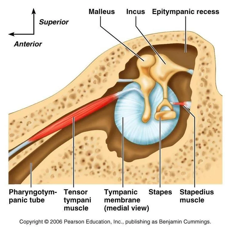
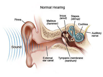
In the process of auditory perception, sound waves from the environment vibrate the tympanic membrane, or the ear drum. These vibrations are passed through a series of three tiny bones collectively called the ossicles. From here, the ossicles cause vibration of the fluid inside the cochlea. Lining the cochlea are cells called hair cells. These hair cells are the ones that send auditory information into the brain, and are highly susceptible to damage by high volume sounds.
In order to protect these delicate hair cells after prolonged exposure to loud sounds, the body reflexively uses muscles to decrease the amplitude of the stimuli that reach the cochlea. In particular, there are two muscles that can constrict which decrease the movement of the ossicles, thus minimizing the damage to the hair cells.
One muscle is called the stapedius muscle. The stapedius muscle inserts into the stapes, the smallest bone in the body and the bone that makes contact with the oval window of the cochlea. The facial nerve (Cranial nerve VII) innervates the stapedius. After long exposure to loud auditory stimuli, the stapedius muscle becomes tightened, which decreases the amplitude of the signal passed onto the cochlea.
The other muscle is the tympani tensor muscle. This muscle inserts into the malleus, the first of the three muscles immediately after the eardrum (tympanic membrane). The mandibular branch of the trigeminal nerve (Cranial nerve V) is responsible for sending signals to the tympani tensor muscle. This muscle can be controlled voluntarily by some people, and is tensed during yawning. The tympani tensor muscle is tightened spontaneously after exposure to loud sounds, and this reflex exists on a time scale of about 40 ms, which may have been sufficient to minimize the damage from loud sounds such as those made during a thunderstorm, but not for more abrupt sounds like gunshots.
Activity of either the stapedius or the tympani tensor muscles can dampen the amplitude of sounds by about 20-30 decibels.
In some cases, an injury to the nerves that innervate these two muscles may result in a hearing disorder called hyperacusis. In hyperacusis, sounds are much louder than they would be in normal conditions. In hyperacusis, people have a higher risk of developing hearing loss due the inability for the stapedius or tympani tensor muscles to decrease the amplitude of sounds. Hyperacusis may occur as a result from traumatic head injury, Lyme disease, or Meniere’s disease. Some drugs may cause hyperacusis, such as LSD or phencyclidine.