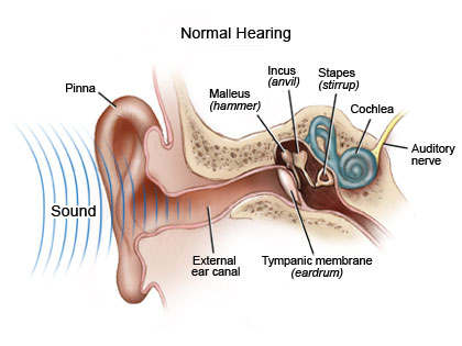
Answer: The auditory nerve is one of the two major branches of the vestibulocochlear nerve (Cranial Nerve VIII). It carries auditory sense information into the brain.
Cranial Nerve VIII, the vestibulocochlear nerve, is a sensory nerve that has two major branches. One of them, the vestibular nerve, carries information from the semicircular canals of the inner ear to the brain. This helps us orient ourselves as to our position in space, whether we are upright, and which way our head is tilting.
The other branch is the auditory nerve, sometimes called the cochlear nerve. It consists of neurons that have their cell bodies in the cochlea that then project into the cochlear nucleus of the medulla in the brain stem. These initial projections are ipsilateral, meaning that auditory information from the left ear projects into the left medulla.
Inside the human cochlea, hair cells are sensitive to a range of frequencies, from 20 Hz up to 20,000 Hz. These cells are responsible for transmitting sound information into electrical signals which the brain can then interpret. The hair cells themselves deflect in response to physical stimulation of the endolymph, the fluid that fills the inside of the cochlea. Changes in orientation of these hair cells are turned into electrical signals, and these are the signals that are sent into the brain via the auditory nerve.
A cochlear implant bypasses the need for the hair cells to be activated. It directly stimulates the auditory nerve, and functions more completely to provide auditory input to the brain as opposed to as traditional hearing aid.
