Answer: The central canal is a fluid-filled space in the spinal cord that has a protective function and allows for nutrient transport.
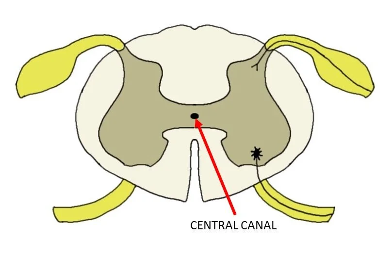
The ventricles in the brain are filled with a high salinity solution called cerebrospinal fluid. This CSF is produced by ependymal cells in the choroid plexus, which lines the ventricles in the brain. CSF functions to increase the buoyancy of the brain, which cushions and supports the brain inside the skull. The ventricular system extends posterior from the brain out of the fourth ventricle into the spinal cord via a structure called the central canal. The boundary between the fourth ventricle and the central canal is called the obex. The central canal is completely continuous with the ventricles, meaning that CSF can flow from the brain into the spinal cord and vice versa.
Moving posteriorly, the central canal forms an enlargement where the CSF fills a slightly larger cavity. This is sometimes called the fifth ventricle or terminal ventricle, and is often found at the level of the first or second lumbar vertebrae, L1 and L2.
Just like the ventricles in the brain, the central canal carries CSF that is rich in nutrients such as glucose. These nutrients are delivered throughout the spinal cord via the central canal.
Learn more neuroanatomy here: